Does COPD Show Up on X-Rays?
August 01, 2023
Content created for the Bezzy community and sponsored by our partners. Learn More

Photography by Charday Penn/Shutterstock
Spirometry is the gold standard for diagnosing chronic obstructive pulmonary disease (COPD). Chest X-rays aren’t useful for diagnosis but can help spot other respiratory issues.
Spirometry is a breathing test that measures how much air you can breathe in and out of your lungs, and how fast. You do this test if your doctor thinks you may have COPD.
But healthcare professionals may also suggest an X-ray when they’re first assessing you for COPD.
According to a 2023 report from the Global Initiative for Chronic Obstructive Lung Disease (GOLD), chest X-rays aren’t sufficient to confirm a COPD diagnosis. But a chest X-ray may be useful in other ways when you’re having breathing issues.
Getting an X-ray can be helpful for detecting or ruling out other conditions that might be causing you to feel out of breath.
Here’s what you need to know about getting chest X-rays when you have (or think you might have) COPD.


What will an X-ray show?
Here’s what a chest X-ray of healthy lungs looks like:
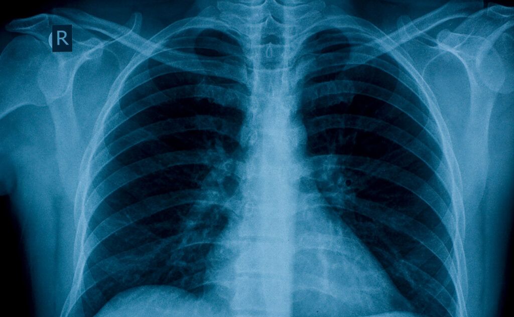
A chest X-ray may show signs of certain respiratory diseases and conditions. However, a chest X-ray isn’t the most accurate way to diagnose COPD.
Why it’s not useful for COPD diagnosis
Researchers in a 2016 study did chest X-rays and spirometry on 183 participants with suspected COPD. They compared the X-ray and spirometry results to see how well the chest X-ray reports matched the gold-standard spirometry diagnosis.
The chest X-ray reports were not very accurate. The researchers found that one-third of people who received a positive diagnosis using the X-ray report didn’t actually have COPD according to spirometry, which is known to provide an accurate diagnosis.
These results suggest that diagnosing COPD using a chest X-ray may lead to misdiagnosis, along with someone receiving the wrong treatment.
Why it may help identify some visual features of COPD
Healthcare professionals may still use a chest X-ray to look for certain features of COPD or other respiratory issues.
A research review in 2010 suggests that certain visuals in a chest X-ray are signs of emphysema, a form of COPD. Visuals include:
- lungs that are easier to see through (less opaque) in the X-ray image
- a flattened diaphragm
- reduced blood vessels visible in some areas
- greater distance from the front to the back of the chest
- wider spaces between the ribs
- a heart silhouette that’s more narrow and vertical
Preparing for a chest X-ray
Getting a chest X-ray is fast and easy.
You don’t have to prepare much ahead of time, but here are some things healthcare professionals may ask you to do right before the X-ray:
- remove jewelry, belts, and other metal items
- take off your glasses
- remove dental appliances
- get undressed and wear a hospital gown
If you’re pregnant, or think you might be, be sure to tell the doctors and technicians before the X-ray. They may take measures to protect the fetus from radiation or decide not to do the X-ray.
What if it’s not COPD?
If you’re having trouble breathing, you may have another condition. A chest X-ray can help identify or exclude it.
A small study in 2009 found that 14% of people who got chest X-rays at COPD evaluations had signs of other potentially treatable causes of breathlessness.
The other potentially treatable causes mentioned were:
- a lower respiratory tract infection, which 51 participants had
- bronchiectasis
- pulmonary fibrosis
- pleural effusion
- left ventricular heart failure
- tuberculosis
The study researchers also found that 11 of the 546 chest X-rays they analyzed showed signs of lung cancer — catching it in the early stages in some cases.
It’s important to note that the study sample size was small and that findings may vary in other studies done in different settings.
If you’re concerned a condition other than COPD may be affecting your breathing, speak with your doctor.
What’s the difference between X-rays and CT scans?
Healthcare professionals often use computerized tomography (CT) scans to investigate the causes of shortness of breath and other respiratory issues.
CT scans use X-rays just like a traditional X-ray procedure. They both direct beams of X-ray light through the body, using detectors on the other side of your body. Your tissues absorb the light in different amounts, so each tissue creates a characteristic pattern in the resulting images.
Here are a few key differences between X-rays and CT scans:
| X-ray | CT scan | |
| Level of detail | less detail | greater detail |
| Number of images produced | two images of your chest — from the back and the side | many images of your body in parallel cross-sections, similar to the slices in a loaf of bread |
| What you do during the scan | you stand or lie down; the technologist may position an arm containing the X-ray tube toward you | you lie on your back on the exam table, which then moves you through a tube for the scan — potentially several times — as the tube rotates around you |
| Number of X-ray beams | a single beam of radiation | several X-ray beams that rotate around you |
| Injection of contrast material | not used | you may receive an injection right before the scan |
| Fasting and medication before the scan | not used | one or both may be required a few hours before the scan |
| Duration | over in seconds; about 15 minutes with prep time | over in seconds; about 30 minutes with prep time |
If you have COPD, a healthcare professional may recommend other scan types as well, such as optical coherence tomography (OCT) and magnetic resonance imaging (MRI).
COPD staging
X-ray imaging doesn’t help healthcare professionals evaluate the stage or severity of COPD.
The 2016 study comparing X-rays with spirometry found that X-ray reports from people with COPD didn’t reliably report differences between those with mild, moderate, and severe COPD.
Not to mention, staging may be on its way out.
The 2023 GOLD report researchers propose a change in terminology for COPD classification that doesn’t use stages. Here are the new classes it suggests:
- Early COPD: Early in the disease process.
- Mild COPD: Low severity of airway obstruction, as measured by spirometry.
- Young COPD: In people ages 20–50.
- Pre-COPD: People of any age with respiratory issues but no airway obstruction.
- PRISm: Individuals with greatly lowered lung capacity (FEV1/FVC ≥0.7 and FEV1 < 80% of the reference capacity).
Takeaway
Chest X-rays may not be a good way of diagnosing COPD, but they can help diagnose or rule out other respiratory issues.
If your doctor thinks you may have COPD, they will probably request that you get spirometry and may recommend getting a CT scan, X-ray, or other type of imaging to help investigate the cause of your symptoms.
Medically reviewed on August 01, 2023
8 Sources
Like the story? React, bookmark, or share below:
Have thoughts or suggestions about this article? Email us at article-feedback@bezzy.com.